

Want to support free content?
Let us know who you are!
Why am I seeing this?
LearnOncology.ca is a free educational content provider. If you want to support our editors (powered by caffeine and no sleep), fill in the following info to let us know who is using the site. We use cookies to prevent this popup from coming up frequently. Continue using the site if you are happy with this.


The following module was designed to supplement learning in the clinic. Please take the time to read through each module by clicking the headings below. By the end of the tutorial, the following objectives should be addressed:
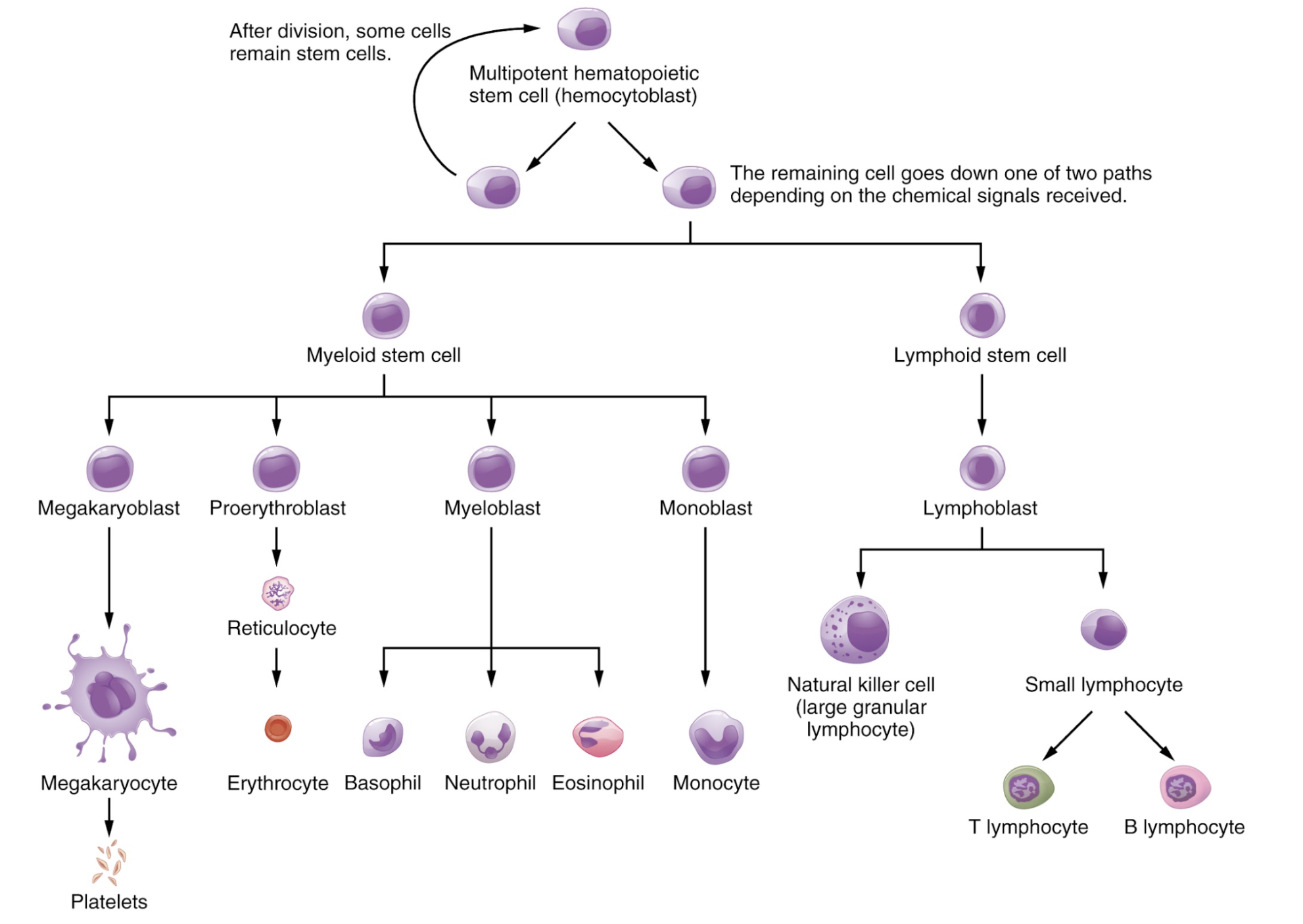
Leukemias are a group of disorders that are defined by an abnormal proliferation of leukocytes, or white blood cells (WBC). Leukocytes, along with platelets and red blood cells (RBCs) arise from hematopoietic stem cells (HSCs) which have the ability to self-renew, or differentiate into various lineages based on chemical stimuli known as hematopoietic growth factors (see Figure 1) [1].
Two major cell lines that arise from HSCs include the myeloid and lymphoid lineages:
Several hormones assist in orderly hematopoiesis including:
Myeloproliferative neoplasms (MPNs) are rare in comparison to solid tumours, and their incidence are inconsistently documented. A recent study from Alberta suggests an incidence rate of 2.05 per 100 000 person-years [2]. While polycythemia vera (PV), essential thrombocythemia (ET) and chronic myeloid leukemia (CML) can occur at any age, primary myelofibrosis (PMF) primarily affects patients older than 60 years of age. PV is the most common of MPNs, while PMF is the least common [3].
Myelodysplastic syndromes (MDS) largely affect older adults, with a mean age at onset of 70 years. MDS is relatively common, with an incidence reaching over 100 per million persons in the general population [4].
Acute myeloid leukemia (AML) accounts for 1.3% of all cancer cases in the United States, and 31% of all new acute leukemias. However, given its aggressive nature, it causes over 60% of all leukemia-related deaths annually [5]. While new treatments have improved mortality and morbidity of AML, long-term survival is still infrequent; Canadian data from 2021 suggests a 5-year net survival rate of 23% [6].
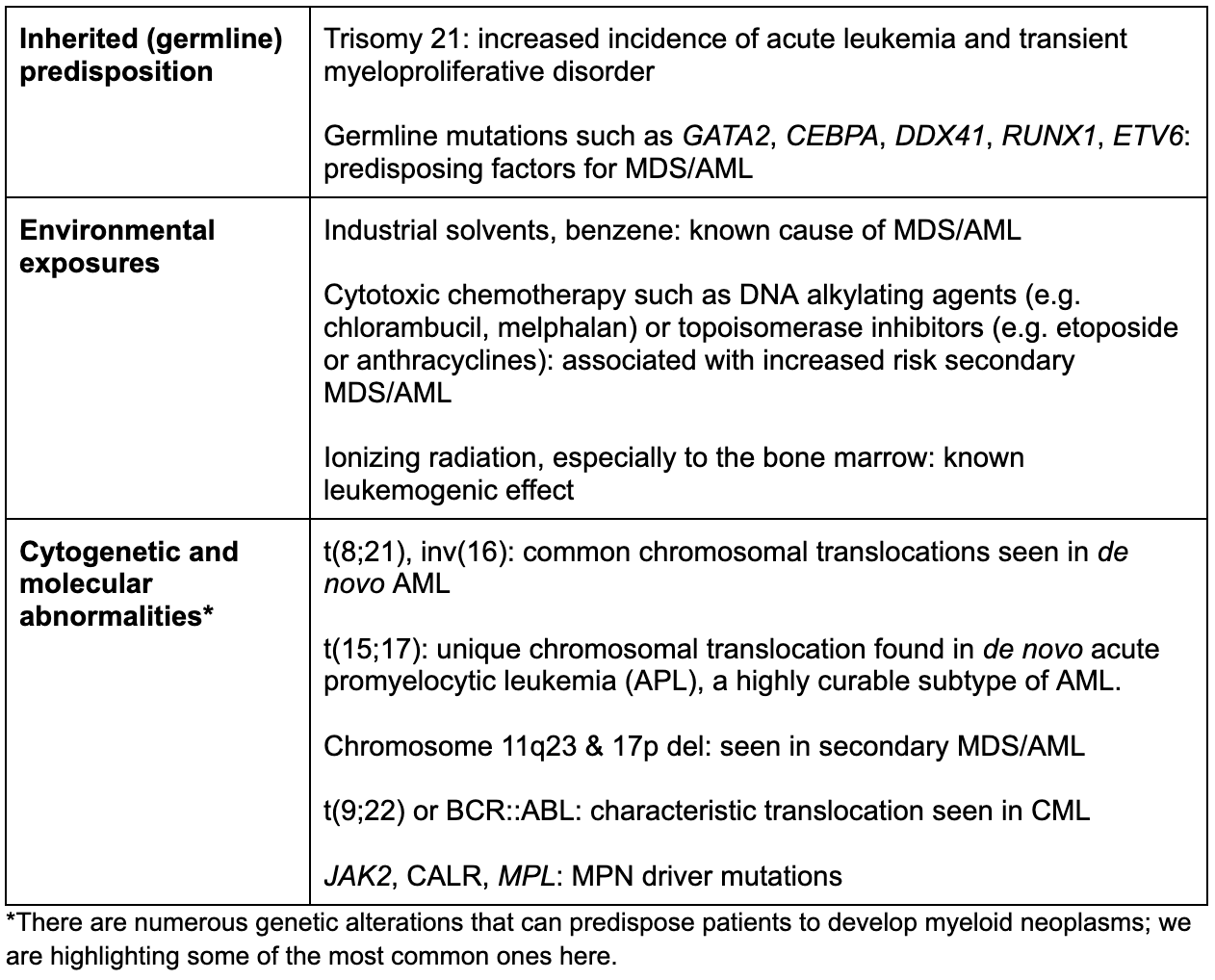
Each myeloid malignancy slightly differs with respect to its specific etiology. In general, cancer is a result of cumulative genetic mutations that can be acquired spontaneously (i.e. de novo) or inherited. Below is a list of common risk factors for developing myeloid malignancies [7].
As hematological malignancies can affect virtually every organ system in the body, symptoms can be quite varied and nonspecific. We can frame the symptoms based on their underlying pathophysiology, while also bearing in mind that many symptoms are multifactorial in nature [8].
Currently, there are no widely accepted screening protocols for myeloid neoplasms.
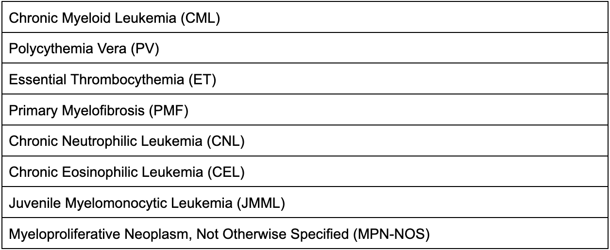
Myeloproliferative neoplasms (MPNs) are a group of disorders characterized by a proliferation of RBCs, granulocytes, and/or platelets without significant dysplasia and with the potential for transformation [10]. The 2022 World Health Organization guidelines have classified MPNs into eight subtypes (see Table 1 below) [10].
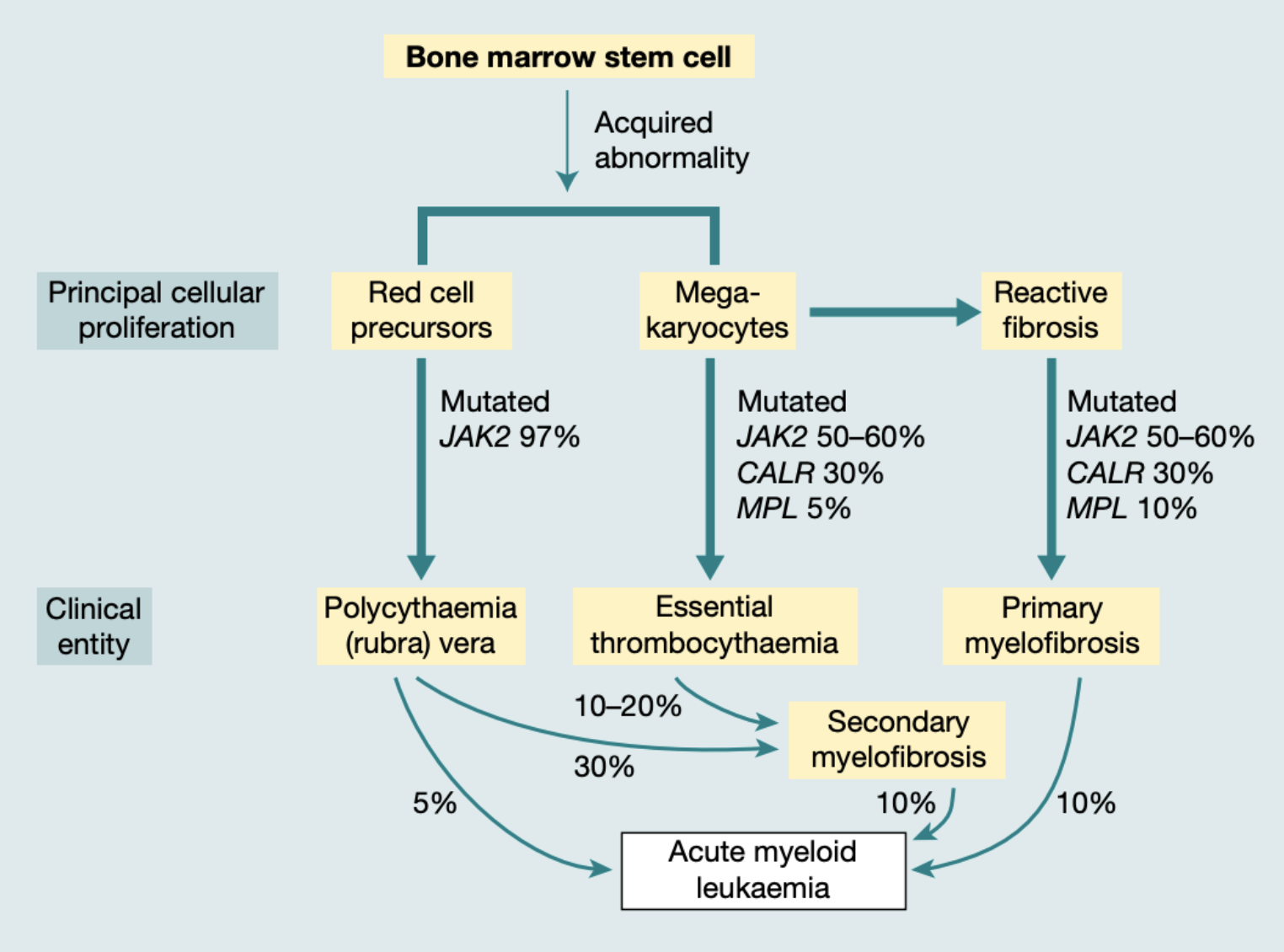
Patients with PV and ET can transform to a secondary myelofibrosis. PV, ET, and PMF can all transform into AML (see Figure 2). Often transformation occurs due to development of new genetic or molecular abnormalities in addition to the initial driver mutation [3].
For the scope of this module, we will briefly discuss the four most common MPNs: PV, ET, PMF and CML.
Polycythemia vera (PV) is defined as an increase in RBC volume as a result of clonal proliferation of a bone marrow stem cell. The JAK2 V617F mutation is seen in about 97% of patients with PV and is presumed to be central to its pathogenesis [11,12]. Patients are typically suspected to have PV after an incidental finding of a high hemoglobin (Hb), but may present with symptoms of hyperviscosity, thrombosis (including unusual sites like hepatic vein thrombosis), or splenomegaly. A few highly suggestive symptoms include 1) aquagenic pruritus, or itchiness provoked by water, and 2) erythromelalgia, which is characterized by burning pain and erythema in the extremities; the pathophysiology of both are unclear, but is thought to be related to mast cell degranulation and platelet-endothelial interactions, respectively [11,13].
PV is diagnosed based on specific criteria including 1) evidence of persistently elevated hemoglobin or hematocrit, 2) bone marrow biopsy confirming increased trilineage hematopoiesis (panmyelosis), 3) presence of JAK2 mutation, and 4) low serum EPO level [11]. Since JAK2 is nearly 99% sensitive for PV, a negative test makes PV unlikely and patients should be worked up for other causes of both primary (i.e. genetic causes) and secondary polycythemia [12]. Secondary polycythemia can be delineated by appropriate EPO production (e.g. high altitude, cardiopulmonary disease, hypoventilation syndromes), or inappropriate (e.g. androgen use, functional renal lesions, ectopic EPO secretion).
Essential thrombocythemia (ET) is characterized by clonal thrombocytosis. Nearly 50-60% of patients present with JAK2 V617F mutations, or an alternate driver mutation that influences JAK2-STAT pathway proliferation and platelet development, such as MPL and CALR [12,14]. Similar to PV, most patients are asymptomatic upon presentation, but common features of ET are thrombosis and hemorrhage due to platelet proliferation and dysfunction [12]. Patients can also present with constitutional symptoms or splenomegaly. Diagnosis of ET can only be made after ruling out reactive causes of thrombocytosis, such as inflammatory or post-surgical states, infection, or iron deficiency [14]. Criteria include 1) thrombocytosis, 2) characteristic megakaryocyte proliferation in the bone marrow, and 3) mutations in JAK2, CALR, or MPL [14].
A diagnosis of primary myelofibrosis (PMF) requires the presence of a clonal marker (e.g. JAK2, MPL or CALR mutation), alongside evidence of significant fibrosis on bone marrow biopsy [12]. These driver mutations are thought to lead to abnormal megakaryocyte proliferation, inflammation and fibroblast overgrowth in the marrow, and extramedullary hematopoiesis.
Clinically, PMF can present as asymptomatic cytopenias in an older adult; however, in contrast to PV and ET, these patients are often symptomatic at presentation. The following findings are common [15]:
Diagnosis of PMF is divided into a pre-fibrotic or overtly fibrotic stage, and is based on bone marrow findings, driver mutations, as well as minor criteria including anemia, leukocytosis, palpable splenomegaly, and an elevated lactate dehydrogenase [15].
While chronic myeloid leukemia (CML) can involve practically any hematopoietic stem cell lineage, it is characterized and diagnosed by the presence of the Philadelphia chromosome. This abnormal chromosome is the result of a translocation between proto-oncogene ABL1 on chromosome 9 and constitutively active BCR gene on chromosome 22. The expression of this BCR::ABL1 oncogene leads to a constantly active tyrosine kinase that inhibits apoptosis and promotes the proliferation of CML cells through aberrant signaling pathways [16,17].
Over half of CML patients are asymptomatic at time of diagnosis, and CML is often first detected from routine bloodwork. However, the most common presenting complaints are related to symptomatic anemia (i.e. fatigue, dyspnea, and pallor) and splenomegaly (i.e. early satiety and abdominal discomfort) [16]. WBCs are typically persistently elevated (>15 x 109/L) with greater percentages of basophils, immature myeloid cells such as myelocytes or metamyelocytes, as well as circulating blast cells. The BCR::ABL1 fusion protein can typically be identified through blood testing and polymerase chain reaction (PCR). Confirmation of a diagnosis of CML is based on morphology on the bone marrow biopsy, along with detection of the Philadelphia chromosome via genetic testing such as karyotyping or fluorescence in situ hybridization (FISH).
CML was originally categorized into three stages: chronic phase (CML-CP), accelerated phase (CML-AP), and blast phase (CML-BP). Around 90% of patients present with CML-CP, which has a slow-growing, indolent course in which there are less than 10% of blasts in the peripheral blood or bone marrow, absence of extramedullary manifestations of leukemia and relatively normal basophil and platelet counts. Conversely, a minority of patients present in CML-BP, also known as “blast crisis” or acute transformation, which is characterized by greater than 20% of blasts in blood or in the marrow [17]. The WHO 2022 classification removed CML-AP and instead uses the category “high-risk CML” to identify patients who are at increased risk of developing advanced CML or transforming. Risk factors include genetic factors, such as additional cytogenetic abnormalities (ACAs) picked up on FISH, treatment resistance, and poor adherence to treatment [18]. With treatment, the 10-year incidence of progression to blast phase disease is 5-6% [16].
Myelodysplastic syndromes (MDS) are characterized by the simultaneous proliferation & destruction of hematopoietic cells, which leads to the signature finding of a hypercellular marrow but peripheral pancytopenia. Most cases of MDS are primary or idiopathic, but there is a category of secondary MDS which includes therapy-related MDS (t-MDS), which is a result of prior chemotherapy or radiotherapy, or MDS arising from mutations or other causes of marrow failure [19].
While there is an inherited predisposition to MDS in patients who are diagnosed at a younger age, the majority of MDS cases have an unknown etiology [20]. Clinical features are variable and non-specific, but typically include complications of reduced cell counts and poorly functioning blood cells, such as recurrent infections, transfusion-dependent anemia, or easy bleeding and bruising. Patients may have cytopenias affecting any number of cell lines, and macrocytosis is common. Abnormal appearing “dysplastic” cells may be seen on a peripheral blood smear, but not universally. Patients with higher risk MDS may also have a small proportion of blast cells in the blood. MDS should be considered in an elderly patient presenting with a macrocytic anemia after nutritional deficiencies, alcohol use, as well as liver and thyroid disease, are ruled out as potential causes. A bone marrow biopsy required for diagnosis typically shows hypercellularity with dysplastic cells and in some cases, excess blast cells in the marrow. Importantly, dysplastic cells can be seen in a variety of conditions including chronic alcohol use, infections such as parvovirus or HIV, and even after chemotherapy or G-CSF treatment. Therefore, these conditions must be ruled out prior to diagnosis [19,20].
In general, acute leukemia is defined by a blast cell percentage of at least 20% in the peripheral blood or bone marrow at presentation. Immunophenotyping of blast cells then allows for diagnosis of either myeloid (AML) or lymphoblastic leukemia (ALL). AML is the most common form of acute leukemia in adults [21]. The WHO 2016 arranges AML into 2 major categories: AML with defining genetic abnormalities, and AML defined by differentiation. Importantly, most AMLs with defining genetic abnormalities may be diagnosed with less than 20% blasts. For all other AMLs with no identifiable driver mutations, the diagnosis is made with evidence of ≥20% blasts in bone marrow or blood [10].
Most cases of AML do not have an identifiable predisposing risk factor or etiology. Known risk factors of developing MDS or AML include cytotoxic chemotherapy, ionizing radiation and benzene exposure, as well as some high-risk genetic conditions (see the “Risk Factors and Etiology” section above). Patients with AML present with nonspecific symptoms or complications from anemia, thrombocytopenia, and neutropenia. Common signs at presentation include fatigue, infections, fever of unknown origin, bleeding, easy bruising, and bone pain. Patients may suffer from hematologic emergencies when they present with AML including febrile neutropenia, leukostasis syndrome, coagulopathy, or tumour lysis syndrome.
On laboratory investigations, cytopenias are common with decreased reticulocytosis due to bone marrow infiltration [22]. On peripheral blood smear, rod-shaped granules in myeloblast cells known as Auer rods are nearly completely specific for AML, and are particularly associated with APL (though Auer rods can also be seen in high risk MDS). Auer rods are representative of myeloid differentiation and are only seen in malignant states [21]. In patients with acute promyelocytic leukemia (APL), a rare subtype of AML, disseminated intravascular coagulation (DIC) is a common yet dangerous presentation that arises from the tendency of leukemic blasts to overactivate the coagulation cascade [22]. APL is a clinical emergency; and early recognition and empiric therapy with all-trans retinoic acid (ATRA) can be life saving.
Specific treatment of all myeloid neoplasms is a complex discussion, as each malignancy has its own management. We will give a high-level overview of the various treatments for each myeloid malignancy; specific details of these can be found in the cited references.
Currently, pharmacological treatment for PV has not proven to be life-prolonging. Therefore, the objective of management is to prevent thrombosis, which is the most dangerous complication of PV. All patients with PV require periodic phlebotomy in order to target a hematocrit of less than 45%, as well as daily, low-dose aspirin to reduce their thrombosis risk. Thereafter, patients are risk stratified based on their age and history of thrombosis; high risk patients receive cytoreductive therapy. The most commonly used cytoreductive agent is hydroxyurea which is superior to phlebotomy alone in high risk patients [11].
ET is treated fairly similarly to PV, with the main goal of preventing thrombotic complications. Treatment recommendations are based on a 4-tiered risk stratification (based on age, thrombosis history, and presence of JAK2 V617F mutation). Typical therapy include antiplatelet agents (ASA) alone in lower risk patients, or combined with cytoreductive agents like hydroxyurea in higher risk patients [14].
The only life-prolonging treatment for PMF, currently, is a stem cell transplant; however, this is associated with a high risk of morbidity and mortality. Therefore, a risk-stratified approach is essential to delineate patients who will benefit from therapy. JAK inhibitors (i.e. ruxolitinib, momelotinib) are commonly prescribed for management of symptoms from PMF, particularly splenomegaly, but this has not been proven to have a survival benefit.
Symptom-directed therapy is important in PMF, as disease progression can significantly impair quality of life. For anemia, erythropoiesis-stimulating agents and androgens can be used along with periodic red cell transfusions. Symptomatic splenomegaly can be treated with hydroxyurea or JAK2 inhibitors.
The mainstay of CML treatment are tyrosine kinase inhibitors (TKIs); TKIs have a significant survival benefit in patients with chronic phase CML. Imatinib is a first-generation TKI, however its use has declined with development of more potent second-generation TKIs (e.g. nilotinib and dasatinib). The specific TKI that is chosen is based on specifics of the disease, prognostic score, and patient-related factors [17]. In most patients, the goal of treatment is to achieve “major molecular remission”, which is defined as a 3-log reduction in BCR::ABL1 transcript reduction from PCR analysis of the peripheral blood or bone marrow. Patients who are not responding optimally to treatment may be developing drug resistance; these patients are then trialed on second-line TKIs [16,17].
CML-BP is difficult to treat, and there is no current universal standard of care. Patients are typically treated with an AML or ALL-directed regime, based on their blast cell immunophenotype, along with a TKI. Patients who achieve remission frequently proceed to allogeneic stem cell transplant, as CML-BP is typically incurable without transplantation [16,17].
The goal of AML treatment depends on the patient and their values, as well as their overall fitness level. If patients are not able to tolerate intensive chemotherapy, the goal of treatment is to improve quality of life and decrease risk of cytopenia-related complications [23].
For patients who are considered fit for intensive therapy, AML treatment is divided into two phases, known as induction and consolidation. The aim of induction is to induce complete remission, which is defined by less than 5% blasts in the bone marrow, with recovery of normal blood counts and improvement in clinical status [21]. The backbone of induction chemotherapy is anthracyclines (idarubicin or daunorubicin), and cytarabine. AML treatment is a rapidly growing field; and many patients are also treated with targeted therapies based on their disease biology (e.g. FLT3 inhibitor).
Once patients achieve complete remission, consolidation therapy is used to eradicate undetectable malignant cells and prevent future relapse. Patients who have favorable-risk AML based on disease biology are typically treated with consolidation chemotherapy alone. Patients with intermediate- or adverse-risk AML are usually offered allogeneic stem cell transplantation once remission is achieved [23].
Due to significant advances in AML research, patients who were previously deemed too elderly or unfit for intensive induction may now be offered a highly potent induction regimen with venetoclax (a BCL-2 inhibitor) and a hypomethylating agent (typically azacitidine). This combination has reduced toxicity compared to conventional chemotherapy and can produce high rates of remission and improved survival compared to historical treatment options [24]. Given these impressive results, this combination is being increasingly studied in younger patients as well.
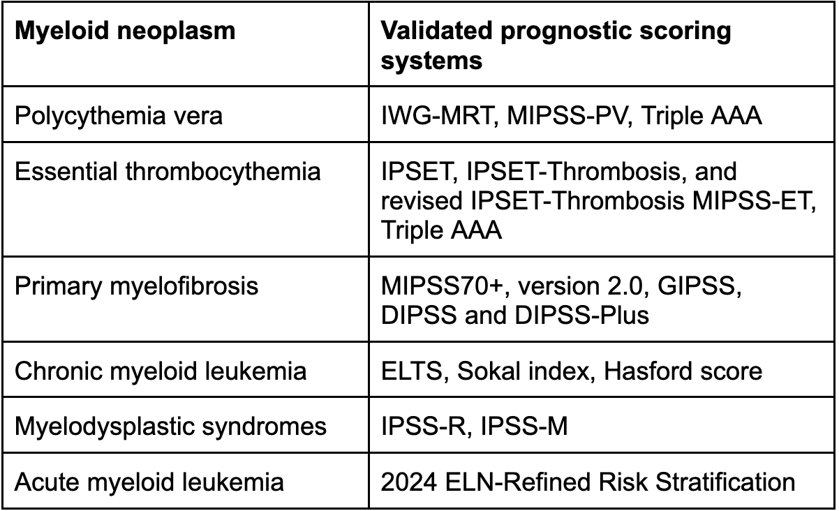
Predicting prognosis in both chronic diseases such as MPNs or MDS, as well as AML, is important in guiding a patient’s treatment course and their goals of care. Most conditions have various validated scoring systems that help clinicians describe the prognosis of patients’ disease to themselves and their families; examples of such scores can be found in Table 3. These scores include patient-related risk factors, such as age at diagnosis, presenting symptoms and history of medical comorbidities, laboratory values such as cell counts, as well as cytogenetic risk factors. The specifics of these scores is not within the scope of this module, but can be found elsewhere [23,25,26].
Use your mouse to click through the slides and answer each question in the text box provided.
Note: This case can be completed on an iPad. To do this download the (free) Articulate Mobile Player for the iPad by clicking here.
